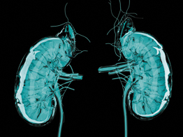
Review: 2012 KDIGO Guidelines Resolved Kidney Disease Controversies, but Uncertainties Remain
A new review article found that Kidney Disease Improving Global Outcomes (KDIGO) guidelines published in 2012 resolved uncertainties and controversies around definitions for acute kidney disease (AKD) and chronic kidney disease (CKD) (JAMA 2015;313:837–46). However, other guidelines are not completely concordant with KDIGO guidelines, and these guidelines as well as KDIGO leave areas of uncertainty in evaluating duration of kidney disease, assessing special populations, determining kidney function in the context of drug development and dosing, and evaluating and predicting CKD progression.
Authors Andrew Levey, Cassandra Becker, and Lesley Inker at Tufts Medical Center in Boston summarized evidence supporting the use of laboratory tests for glomerular filtration rate (GFR) and albuminuria to detect and stage acute kidney injury (AKI), AKD, and CKD.
In addition to reviewing KDIGO and other guidelines, Levey and his colleagues searched the scientific literature for cystatin C-based equations for estimated GFR (eGFR) published after KDIGO 2012. The other guidelines included the National Kidney Foundation Kidney Disease Outcomes Quality Initiative (NKF-KDOQI), a Canadian Society of Nephrology (CSN) commentary, National Institute for Clinical Excellence (NICE) guidelines, and Kidney Health Australia Caring for Australasians With Renal Impairment (KHA-CARI).
The authors concluded that KDIGO harmonized definitions and staging systems for AKI published before KDIGO 2012. These definitions and staging systems defined AKI based on increased serum creatinine or urine volume. NKF-KDOQI, CSN, NICE, and KHA-CARI all concur with the KDIGO AKI definition and staging, though, some, like NKF-KDOQI, differ slightly with KDIGO oliguria criteria.
The authors found that several new eGFR equations have been reported since KDIGO 2012, but none appear to be more accurate in North America, Europe, and Australia than the 2009 Chronic Kidney Disease Epidemiology Collaboration (CKD-EPI) equation. Since eGFR based on creatinine may be less accurate for clinical decision-making during AKI, the authors recommended confirmatory eGFR based on serum cystatin C, with or without accompanying serum creatinine (eGFRcr-cys; eGFRcys). It might be preferable to rely on eGFRcys rather than eGFRcr-cys in patients likely to have inaccurate eGFRcr due to non-GFR factors.
Measuring the albumin-to-creatinine ratio
in an untimed specimen is the preferred method for assessing albuminuria from a spot urine sample rather than a timed urine collection.
No Xanthochromia, Low RBC
Count in CSF Rules Out Aneurysmal Subarachnoid Hemorrhage
No xanthochromia and red blood cell (RBC) count <2000 x 106/L in cerebrospinal fluid (CSF) reasonably rules out aneurysmal subarachnoid hemorrhage in patients presenting with acute headache (BMJ 2015;350:h568). Most patients who meet this threshold require no further work-up and aneurysmal subarachnoid hemorrhage reasonably can be excluded as the cause of their headache, according to the authors.
The findings were from an observational study of 1,739 patients age 15 or older who presented with acute headache to 12 Canadian emergency departments.
According to the authors, computed tomography has 100% sensitivity for subarachnoid hemorrhage when it is performed within 6 hours of acute headache onset. However, CT’s sensitivity declines to 85.7% when conducted more than 6 hours after symptom onset. Traumatic spinal taps causing blood in CSF occurs in up to 30% of spinal taps, making it difficult to determine whether blood in CSF is from a tap itself or from a subarachnoid hemorrhage. Xanthochromia also is a sign of subarachnoid hemorrhage.
The investigators found that the RBC threshold of <2000 x 106/L in CSF and no xanthochromia excluded a diagnosis of subarachnoid hemorrhage with 100% sensitivity and 91.2% specificity
Vendor-Dependent Issues Contribute to Discordance
in Assigning Antibody Specificity
An analysis of data from the American Society for Histocompatability and Immunogenetics (ASHI) proficiency testing (PT) program suggests that vendor-dependent issues may be behind discordance in assigning antibody specificity (Transpl Immunol 2015;32:1–8). Insufficient concordance and standardization in antibody testing could have practical implications for organ allocation and organ sharing programs, according to the authors.
While anti-HLA antibodies have long been recognized as crucially important in organ transplantation, discussions are still ongoing about the optimal method for detecting them. The investigators accessed data from seven ASHI PT surveys undertaken by an average of 124 laboratories. As part of the PT survey, participating labs were asked to identify: the methods used and source of their testing kits; antibody screening/detection results for each serum or method; and antibody identification results with a positive or negative assignment of each possible antibody specificity.
In analyzing the PT data, the authors found that although 94% of labs used single antigen Luminex beads (SALB), assignment of an average of 10 antibody specificities were discordant for each serum. The assignment disagreement existed for both common and uncommon antigens.
While the authors emphasized that these variances could be due to differences in testing procedures or in result interpretation, they also suggested vendor dependent factors could be at work. For each positive serum sample, an average of 15 antibody assignments was significantly different between users of the two available SALB kits, and intra-vendor concordance was higher than inter-vendor concordance. “Some of the ‘vendor dependent’ assignments are clearly due to differences in the antigen panel represented on the SALB provided by the vendor,” they wrote.
High NPV for Negative Troponin Results in Pulmonary Embolism Work-up
New research supports the use of a contemporary cardiac troponin I (cTn) assay as a risk stratification tool in the work-up of suspected acute pulmonary embolism (PE) (Chest 2015;147:685–94). The findings show that cTn adds incremental prognostic value to clinical, electrocardiographic, and radiographic data and provides “excellent prognostic negative predictive value,” thereby aiding clinicians in identifying patients at low risk for adverse events who might be considered for out-of-hospital management of acute PE, according to the authors.
The retrospective study involved 298 consecutive patients with confirmed acute PE, who had cTn measured by VITROS Troponin I ES Assay, which has a lower limit of detection of 0.012 ng/mL and a 10% coefficient of variation at the 99th percentile upper reference limit of 0.034 ng/mL. The investigators hypothesized that undetectable cTn values by this method would predict very low in-hospital adverse event rates.
Overall, 55% of patients were cTn-positive, while 45% were cTn-negative. No deaths or hard events such as death or cardiopulmonary resuscitation occurred in the cTn-negative patients during the median 5-day in-hospital follow-up period, and in comparison to cTn-positive patients, cTn-negative patients experienced a lower rate of soft events like intensive care unit admission and had higher survival rates.