blueshot / Getty Images
The American Urological Association (AUA) is now discouraging sole use of dipstick tests to diagnose microhematuria and suggests categorizing patients into three risk categories following diagnosis to determine next steps. A multidisciplinary panel revised AUA’s guidelines on microhematuria, which address diagnosis and evaluation of this condition as well as follow-up procedures.
“The goal of the new guideline is to provide a risk-stratified approach to hematuria evaluation based on a patient’s risk factors for urinary tract cancer,” said Daniel Barocas, MD, co-chair of the guideline panel and associate professor of urology at Vanderbilt University, in a statement. “We crafted the guideline with the intention of reducing the intensity of evaluation in those at low risk for malignancy, while preserving the diagnostic sensitivity of evaluation in those at higher risk.”
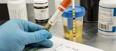
To define/diagnose this condition, the guideline authors are advising that clinicians do a formal microscopic evaluation to follow up on any positive urine dipstick test. “Urine dipstick testing detects the peroxidase activity of hemoglobin using benzidine but does not correlate perfectly with microscopic evaluation,” explained the authors. Certain factors can lead to false-positive dipstick results, including dehydration, exercise, menstrual blood, myoglobinuria, or povidone-iodine (Betadine). The guidelines cite several studies in which the dipstick test produced inconsistent urinalysis (UA) results.
Clinicians should define microhematuria as the microscopic evaluation of a single urine specimen that reads ≥3 red blood cells per high-power field (RBC/HPF). A recent study of more than 46,000 patients found that the highest sensitivity for identifying bladder cancer and lowest negative likelihood ratio existed at ≥3 to 10 RBC/HPF.
In their initial evaluation of patients with microhematuria, clinicians should dispense with urine cytology or urine-based tumor markers in favor of a risk-stratified approach classifying patients as low, intermediate, or high risk for genitourinary malignancy based on certain parameters. Not enough data exist to support the clinical benefits of these markers in patients undergoing cystoscopy to identify bladder cancer, according to the authors.
“Clinicians may obtain urine cytology for patients with persistent microhematuria after a negative work-up who have irritative voiding symptoms or risk factors for carcinoma in situ,” the panel advised.
Looking forward, gaps in knowledge in this area warrant further exploration of additional approaches and technologies. This includes new automated instruments that operate on flow cytometry or digitized microscopy to perform UA. “These machines may not correlate directly with traditional urine microscopy, and thus it will be important to determine if the threshold of 3 RBC/HPF used in the guideline will be an equivalent predictor of risk when these new technologies are used in evaluation,” wrote the authors.
Urinary biomarkers may also serve as a useful tool in tailoring evaluation approaches in microhematuria patients. “Urothelial cancers are in contact with the urine, and this fact has been utilized to evaluate the differential expression of proteins, RNA, DNA, and changes in methylation and cells among patients with malignant and benign conditions,” the authors noted.
Multiple candidate markers are being evaluated as aids in detecting bladder cancer in patients with hematuria. Guideline authors mentioned a prospective trial underway that will randomize participants based on clinical risk and urinary marker status.