Case: The patient is a 30 year old male. CBC showed a normal hemoglobin concentration, hematocrit and mean corpuscular volume (MCV). Hemoglobin electrophoresis was ordered because the patient had been told that he had sickle cell trait. On alkaline gel electrophoresis, the patient's sample is in the second complete lane (see below).
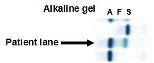
Alkaline gel electrophoresis showed 2 major hemoglobins: an anodal peak of 71.5% and a cathodal peak of 27.4%. Hemoglobin A2 was not elevated. The anodal peak migrated in the normal location of hemoglobin A whereas the cathodal peak migrated in the normal location for hemoglobin S. Is this sickle cell trait? How should you proceed?
Response:
In the case of sickle cell trait, hemoglobin S is confirmed by either a positive sickle solubility test (e.g., mixing the reducing agent sodium hydrosulfite with blood inducing sickling in cells containing hemoglobin S) or by performing an acid gel electrophoresis where hemoglobin S migrates separately from hemoglobin A.
In this case, the sickle solubility test was negative ruling against the non-A peak being hemoglobin S. As well, the A to S ratio in sickle cell trait is ~60:40 and the ratio in this patient is higher than 60:40 (i.e., ~70:30).
Acid gel electrophorsis confirmed that the non-A peak was not hemoglobin S as the acid gel did not reveal an S peak, but did reveal a single large peak migrating in the location of hemoglobin A (middle lane on the acid gel below). The differential diagnosis of this alkaline gel second peak are hemoglobins D, G-Philadelphia, Lepore and Korle-Bu.
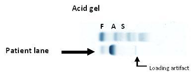
Because hemoglobin Lepore produces a thalassemic phenotype, A/Lepore is unlikely. Therefore this patient could be heterozygous for A and D, A and G, or A and Korle-Bu. Hemoglobin D in the heterozygous state with A (i.e., AD) or homozygous state (i.e., DD) is benign; however, heterozygosity of D with S (i.e., SD), produces a sickle cell anemia-like-phenotype that can produce crises. In order to differentiate D from G from Korle-Bu, molecular analysis was recommended.